13TH INTERNATIONAL CONFERENCE ON MEDICAL INFORMATION PROCESSING AND ANALYSIS (SIPAIM), 2017
Abstract
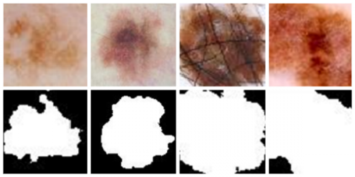
Melanoma skin cancer diagnosis can be challenging due to the similarities of the early stage symptoms with regular moles. Standardized visual parameters can be determined and characterized to suspect a melanoma cancer type. The automation of this diagnosis could have an impact in the medical field by providing a tool to support the specialists with high accuracy. The objective of this study is to develop an algorithm trained to distinguish a highly probable melanoma from a non-dangerous mole by the segmentation and classification of dermoscopic mole images. We evaluate our approach on the dataset provided by the International Skin Imaging Collaboration used in the International Challenge Skin Lesion Analysis Towards Melanoma Detection. For the segmentation task, we apply a preprocessing algorithm and use Otsu’s thresholding in the best performing color space; the average Jaccard Index in the test dataset is 70.05%. For the subsequent classification stage, we use joint histograms in the YCbCr color space, a RBF Gaussian SVM trained with five features concerning circularity and irregularity of the segmented lesion, and the Gray Level Co-occurrence matrix features for texture analysis. These features are combined to obtain an Average Classification Accuracy of 63.3% in the test dataset.